Working Time
- Mon-Thu 08:00 - 20:00
Friday 07:00 - 22:00
Saturday 08:00 - 18:00
Contact Info
-
Phone: 1-800-267-0000
1-800-267-0001 - info@dentco.net
Ask the Experts
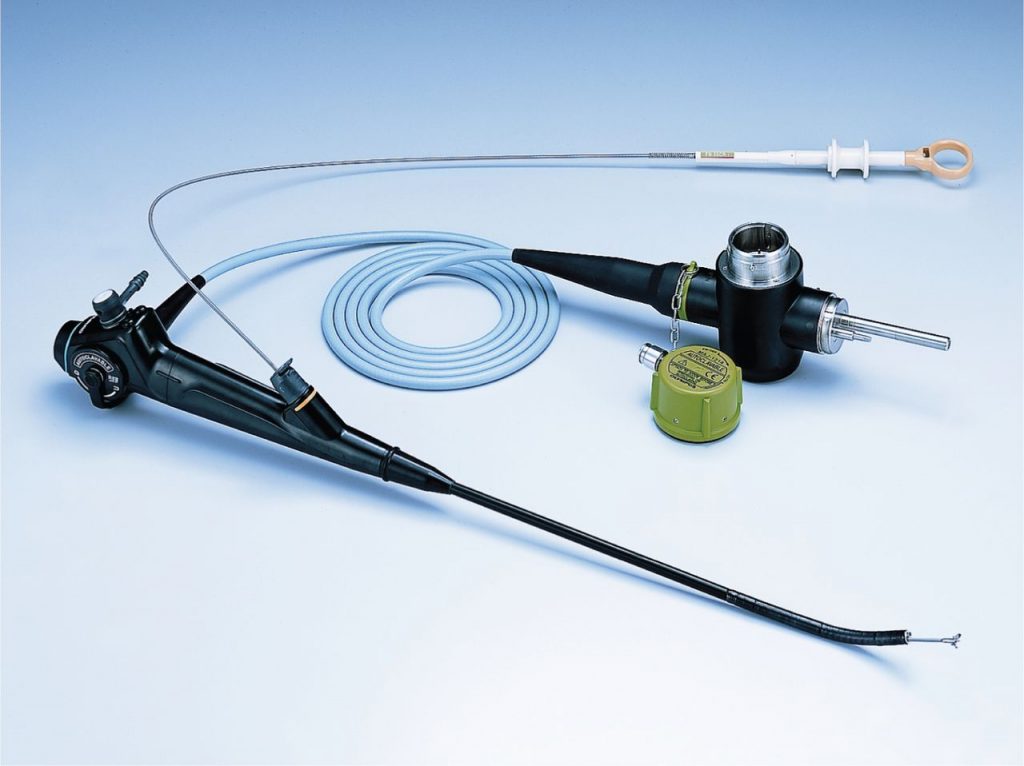
Medical Thoracoscopy

Medical Thoracoscopy (pleuroscopy) involves passage of a camera through the chest wall for direct visualization of the pleura. Thoracoscopy is performed for diagnostic as well as therapeutic purposes. It is performed by pulmonologists. Medical thoracoscopy is most commonly used for pleural fluid drainage, parietal pleural biopsy, and pleurodesis. The most frequent indication for diagnostic thoracoscopy is pleural effusion. Thoracoscopy in spontaneous pneumothorax can identify the cause of the pneumothorax. Medical thoracoscopy is most commonly performed when multiple attempts at thoracentesis (typically two) have failed to achieve a diagnosis in patients with a recurrent exudative pleural effusion.
The thorcoscopy is performed usually under local anaesthesia with conscious sedation, but rarely general anaesthesia may be required. The procedure involves introduction of the instrument in pleural space through a small hole in the chest wall. A chest tube is left in the pleural space after the procedure for the duration required.
However, just as with other types of prodedures, there are inherent potential risks of this procedure. The incidence of major and minor complications associated with thoracoscopy are 1.9% and 5.5%, respectively. The complications include prolonged air leak, hemorrhage, subcutaneous emphysema, postoperative fever, empyema, wound infection, cardiac arrhythmias, reexpansion pulmonary edema, hypotension and seeding of chest wall in patients with mesothelioma. The mortality rate associated with thoracoscopy performed is 0.09% or 1 death in 8000 procedures.

